PhD student Lauren Ryall-Waite shares her visit to Barts Pathology Museum.
My research in the history of surgical emotion is concerned with the use and display of surgically anatomised human remains. I am particularly interested in how they are displayed and in how we engage with such content in museum spaces.
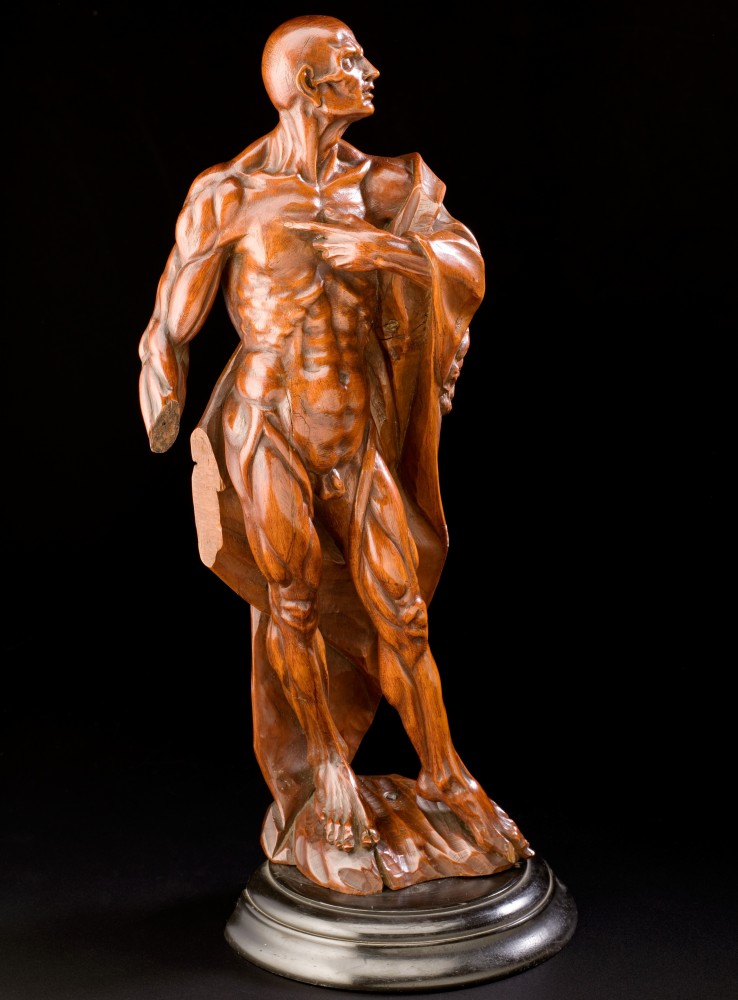
The continued display of human remains in museum spaces has long been a contested topic, and medical museums with pathology collections have been a part of this conversation, particularly when considering 'famous' specimens, such as the Irish Giant.[1] I never miss an opportunity to visit body parts in jars, and I was delighted to see Barts were having an open day to allow anyone to visit the impressive collection.
I must here apologise for the lack of photography in this piece; Barts has a no photography policy, which is very well explored on their website. This is a common theme within museums and echoes the Human Tissue Authority’s respectful stance on the display and usage of human remains in the public eye. I would like to encourage readers to visit the Barts Pathology Museum website, where you can catch glimpses of the building and exhibitions. While it might not always be seen as wise to use one’s imagination too much when considering a collection of wet anatomical specimens, it may assist with the following description of the experience.
If you have not had the chance to visit this unique space yet, it is housed in the impressive historic grounds of St Bartholomew’s Hospital. Because the pathology collection isn’t open to the public except for special events, the collection is not housed within St Bartholomew’s Hospital Museum but in another part of the hospital building. This location adds to the authentic 'behind the scenes' feeling you get as you climb the stairs to reach the pathology collections.
The main room holding the 5000 pathological specimens is light and airy, with jars stretching up towards the ceiling on mezzanine levels connected by an impressive spiral staircase. Your eye is immediately drawn upwards to the light, with the glass ceiling providing ample illumination. Light is generally considered the enemy in museum spaces, with sunlight known for causing damage to historical collections; however, the specimens are well protected from this by their careful placing on shelving which covers most of the available wall space. The result is a bright and lofty space which welcomes visitors in and certainly challenges the stereotype of historic pathology collections being kept in gloomy museum cases in dark rooms.
The specimens themselves are utterly fascinating. Card catalogues attached to the edges of display cases provide information on the specific body parts contained within the jars. The bright and airy feel of the room is no doubt a relief to anyone who may have sensitivities to some of the subject matter on display. The audience here on the open day clearly knew what to expect and were avidly reading the cards and exploring the content with no apparent discomfort at the subject matter. The open shelving design gives visitors a very close-up view of the specimens, and the lack of glass-on-glass which you often experience in medical museums means that you can appreciate all of the minute details of the bodily parts on display without the usual glare. The result is a surprisingly up close and personal experience with human remains in all their glory.
Potted Histories
The brief descriptions provided with the jars are capable of giving us a glimpse into the medical reality of the past. One pot contained the amputated feet of a nineteen-year-old woman which were removed due to frostbite, most likely from sleeping in the cold. This specimen was dated to 1830, meaning the patient would not have had much in the way of anaesthesia or pain relief for the amputation procedure. There is no indication if the young woman survived the procedure or what additional medical issues she may have had; the specimen tells just the medical story, not the personal one.
The museum explores historic occupational hazards, with a collection of severed thumbs and attached tendons which had been ripped out by working machinery in the nineteenth century; a risk common among the factories of the industrial revolution known as avulsion fractures. These rather gory injuries seemed to have moderately positive outcomes in comparison to some of the other afflictions on display, with labels detailing that people went back to work without the difficulty you might imagine after losing a thumb. Phossy jaw and chimney sweep cancer, also known as 'Soot Wart', are also present in the displays. These severe conditions are accompanied by interpretation detailing the working conditions which caused them.
The museum also contains some non-human specimens which I personally found far more discomforting than any of the human remains on display. I encourage people averse to many-legged creatures to avoid this section possibly; the potted giant centipede and preserved horse stomach infected with botfly larvae were particularly memorable.
One display which was very popular on this open day was the intriguing Arnold and Sons cabinet full of things surgeons have removed from people’s bodies. A patient in the 1930s who was in the RAF was using an anti-aircraft shell casing to treat his haemorrhoid problem, presumably inserted in the rectum as a way of preventing prolapses. This slightly uncomfortable DIY answer to a medical problem is one of many unusual things removed from bodies which have been retained in this collection for medical history. Any puns aside, the museum presents such cases in an informative and empathetic manner and encourages respectful responses from its audience. Staff who work with the collections are on hand during the open day to offer assistance in understanding the specimens, ensuring the atmosphere in the museum does not descend into gruesome fascination.
The only frustration I find after visiting pathology collections is that the histories they contain, much like the specimens themselves, are potted. It is probably what makes them so fascinating, the tantalisingly small amount of information we discover of the people represented within the jars makes us want to know more.
Sharing pathological collections with a broader, non-specialist audience has seen a re-focus of interpretation in museum spaces to explore wider issues than purely the history of medicine. Pathological collections naturally lend themselves to discussing health and wellbeing topics with audiences, and many medical museum collections are active in addressing this agenda.[2] The collection at Barts is ideally placed to have such discussions, bringing together the profoundly personal histories present in the specimens along with the modern surgical practices and health interventions.[3]
It is the potential in collections of surgically anatomised human remains like the one at Barts which I find so exciting. The histories surrounding specimens are inherently emotional, drawing on the tragedies of disease as well as the exciting potential of medical and surgical interventions. Museums can, and many museum professionals argue, should, be emotional spaces where people can express their feelings around the past and contextualise the present and future.[4] Pathology collections allow us to address our feelings surrounding disease, death, surgery and medical intervention, as well as our own health and mortality. Barts is a fascinating space where these human experiences meet with the surgical profession, allowing visitors to discover these stories for themselves. It is a visit I strongly recommend if you have the opportunity.
For more information on the image of St Bartholomew click here.
For more information about phossy jaw click here and here.
[1] See Museums Association News https://www.museumsassociation.org/museums-journal/news/20062018-hunterian-to-consider-release-of-irish-giant-skeleton for recent discussions on removing the Irish Giant from display.
[2] See Helen Chatterjee and Guy Noble, Museums, Health and Wellbeing (Routledge, 2013), p.34 for discussion around medical museums contributing to the health and wellbeing agenda.
[3] See https://www.qmul.ac.uk/pathologymuseum/about/ for links to past and future events.
[4] See http://nckultur.org/wp-content/uploads/2015/03/David-Fleming_National-Museums-Liverpool_-The-Emotional-museum-.pdf for exploration of Liverpool Museums as emotional spaces by David Flemming.
Image: Robert Liston operating. Henry Board (c. 1912). (Wellcome Images)